|
Cladosporiaceae
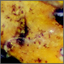
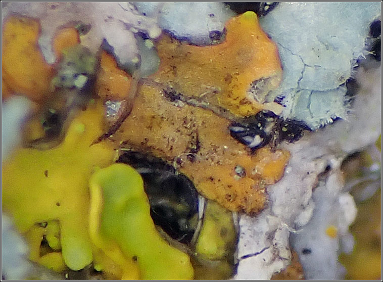
Cladosporium licheniphilum Lichenicolous fungus. Photographs on Xanthoria parietina Probably under-recorded on apothecia and thalli of Xanthoria parietina A dark reddish-brown spiky layer on the host is caused by erect or slightly curved conidiophores, either solitary or in small, loose tufts. They arise from brown, septate hyphae deeply immersed in the host tissue, and produce lemon-shaped or ellipsoid-subcylindrical conidia at their tips. Conidia pale brownish, 0-1 septate, aseptate conidia 3.5-8 x 3-5 µm, the septate conidia 7-13 x 5-7 µm. Micrographs below. Similar: Other Cladosporium species (a large and incompletely understood genus). Gonatophragmium lichenophilum, green-brown, furry coating |
Micrograph
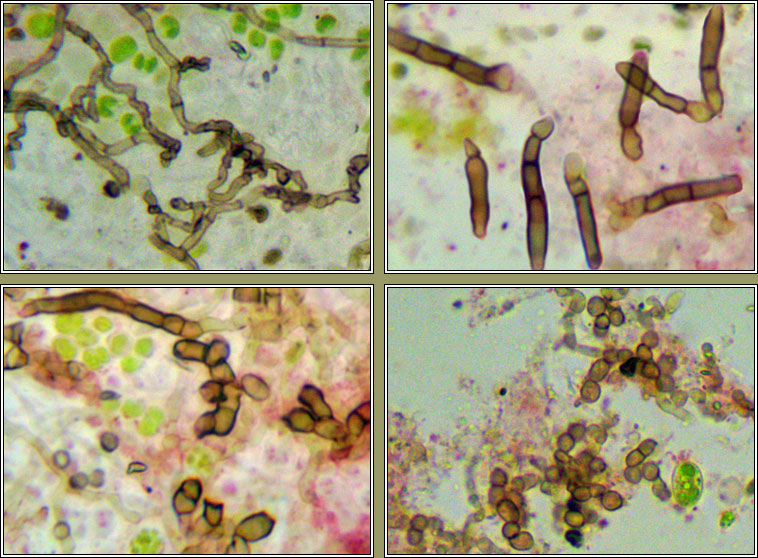
Cladosporium licheniphilum
Photographs: Kingston Lacy, Dorset. July 2023